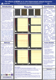
During weeks three and four of my summer research project, I engaged in a meticulously planned and executed investigation involving pharmacological treatments on plated hippocampal cells utilizing MDMA. The initial focus of these weeks revolved around the essential preparatory steps for plating the multi-electrode arrays (MEAs). Over the course of the first few days of each week, I undertook a series of procedures to ensure optimal conditions for the subsequent experiments. To enhance the hydrophilicity of the MEAs, I immersed them in water, as neurons generally exhibit a tendency to reject glass surfaces, impeding their adhesion. Consequently, we coated the MEA surfaces with two types of adhesion molecules, namely PDL and laminin, to promote cell attachment. After allowing the adhesion molecules to soak, plating cell media was applied to the MEAs, which were then left to soak for an additional day while the dissection took place.
On both Thursdays, a critical component of the experiment entailed the dissection of hippocampi from E18 mice. The dissected tissue underwent a process involving trypsinization and trituration, resulting in a solution of cells. The purpose behind conducting weekly dissections was to ensure a consistent supply of mature neurons that could be reliably detected using our specialized hardware. Consequently, the neurons obtained during the initial week had been developing for a duration of 15 days, rendering them suitable for testing the effects of MDMA. In order to facilitate this, a 50 uM solution of MDMA was prepared and combined with maintenance media. This MDMA solution was applied to one of the MEAs, while the other MEA served as a control. On DIV 15, three sets of recordings were made: a control recording, a pre-MDMA baseline recording, and a post-MDMA recording. Visual observations indicated a discernible increase in the number of spike bursts, as well as a notable reduction in the time intervals between bursts, following the application of MDMA.
In the subsequent week, recordings were conducted on both the control and modulated MEAs on DIV 18 to assess the longer-term effects of MDMA. Interestingly, the effects of MDMA were found to be transient in nature, with no immediate or overtly enduring impact on the neurons. Recording sessions were extended to DIV 18, 19, 20, and 21 before disposing of the neurons and proceeding with the cleaning of the MEAs.
The data acquired during these weeks serves as the fundamental basis for my forthcoming presentation. The next phase involves the processing of this data through customized software to generate raster plots, which will effectively quantify the changes associated with MDMA.
Hello, my name is Omar Sbaih, and I am a rising junior in the College of Arts and Sciences. I am majoring in neurobiology with a minor in philosophy and cognitive science. As a Laidlaw Scholar, I am eager to engage with and learn from my fellow scholars, embracing the opportunity to collaborate and share knowledge. I believe that the exchange of ideas and perspectives is vital in expanding our understanding of the world. By actively participating in this community, I hope to gain insights from diverse academic backgrounds and contribute my own unique insights to the collective learning experience.
With access to such a profoundly diverse network, I am driven to explore the underlying principles of human cognition and consciousness through the lens of many different disciplines. I am particularly interested in investigating the ethical implications and societal impact of advancements in neuroscience, particularly the advancement of new pharmacological treatments for cognitive impairments.
Please sign in
If you are a registered user on Laidlaw Scholars Network, please sign in