Week two was primarily dedicated to testing procedures. As with the previous week, significant preparation was required to establish an optimal growth environment for the cells. In anticipation of the dissection scheduled for Thursday, we prepared three regular microelectrode arrays (MEAs) and three partitioned MEAs, employing the methods outlined in last week's post. Subsequently, I conducted the dissection of three embryonic mouse hippocampi, utilizing biological and physical treatments to dissociate and digest the cells for subsequent plating.
On Friday, we obtained our initial set of results, which elicited a mixed response. On one hand, we observed the healthy state of our neurons and the emergence of synapses. However, we were disappointed to discover that the partitioned MEAs provided by the manufacturer incorporated a material with lower conductivity compared to the regular MEAs, leading to the display of only field potential waves. Although disheartening, this test demonstrated that the electrodes in the partitioned MEAs are functional but require a more conductive material to yield satisfactory results.
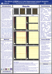
In light of these findings regarding the partitioned MEAs, we redirected our efforts towards a new, yet related avenue of investigation. The laboratory focused on exploring the electrophysiology of serotonin. To achieve this objective, we turned to a well-known pharmacological treatment known to alter the serotonergic nature of neuronal networks: 3,4-Methylenedioxymethamphetamine (MDMA). Limited prior research has been conducted on network electrophysiology in the presence of MDMA, thereby presenting an exciting new frontier for exploration. Over the upcoming weeks, our plan is to formulate MDMA-cell culture media solutions and modulate our established healthy networks to observe any changes in network activity and synchronization.
Hello, my name is Omar Sbaih, and I am a rising junior in the College of Arts and Sciences. I am majoring in neurobiology with a minor in philosophy and cognitive science. As a Laidlaw Scholar, I am eager to engage with and learn from my fellow scholars, embracing the opportunity to collaborate and share knowledge. I believe that the exchange of ideas and perspectives is vital in expanding our understanding of the world. By actively participating in this community, I hope to gain insights from diverse academic backgrounds and contribute my own unique insights to the collective learning experience.
With access to such a profoundly diverse network, I am driven to explore the underlying principles of human cognition and consciousness through the lens of many different disciplines. I am particularly interested in investigating the ethical implications and societal impact of advancements in neuroscience, particularly the advancement of new pharmacological treatments for cognitive impairments.
Please sign in
If you are a registered user on Laidlaw Scholars Network, please sign in