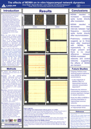
The nature of my summer project entails a significant focus on preparation, as ensuring the smooth progression of subsequent weeks is of utmost importance. Cultivating cells on electrodes embedded in glass presents a challenging task, as neurons typically struggle to thrive outside their aqueous and nutrient-rich environment in the hippocampus. Consequently, my efforts in the initial week were primarily dedicated to preparing the microelectrode arrays (MEAs) for cell plating and assisting in the dissection process.
To enhance the affinity of neurons to the MEAs, a hydrophilic surface was established by immersing them in water. Since neurons generally exhibit resistance to glass adhesion, we applied a coating of two types of adhesion molecules, namely PDL and laminin, to the surface of the MEAs. Subsequently, we allowed the MEAs to soak in cell plating media for an additional day while conducting the dissection process, after which embryonic mice hippocampi were dissected and dissociated using trypsin. The resulting cell population was subjected to ten triterations utilizing a flame-treated pipette tip, yielding viable cells. These cells were then plated onto the MEAs, and we now await the outcome in the upcoming week to determine their survival.
The forthcoming week's plans heavily rely on the viability of the cells. Should the cells successfully survive, our next steps will involve seeking signals through an amplifier and potentially introducing modulation with nicotine. Conversely, if the cells do not survive, we will prepare additional MEAs for dissection and replicate the procedures undertaken in the previous week.
Hello, my name is Omar Sbaih, and I am a rising junior in the College of Arts and Sciences. I am majoring in neurobiology with a minor in philosophy and cognitive science. As a Laidlaw Scholar, I am eager to engage with and learn from my fellow scholars, embracing the opportunity to collaborate and share knowledge. I believe that the exchange of ideas and perspectives is vital in expanding our understanding of the world. By actively participating in this community, I hope to gain insights from diverse academic backgrounds and contribute my own unique insights to the collective learning experience.
With access to such a profoundly diverse network, I am driven to explore the underlying principles of human cognition and consciousness through the lens of many different disciplines. I am particularly interested in investigating the ethical implications and societal impact of advancements in neuroscience, particularly the advancement of new pharmacological treatments for cognitive impairments.
Please sign in
If you are a registered user on Laidlaw Scholars Network, please sign in