Mechanical Quantification of Fibrosis in 2D & 3D Models using Atomic Force Microscopy
Preview
I spent my industrial placement year at GSK (GlaxoSmithKline), a global biopharmaceutical company
known for its focus on innovative medicines and research across a range of therapeutic areas, including
respiratory conditions, oncology and infectious diseases. Within GSK, I was part of the Respiratory
Immunology Inflammation Biology Unit (RIIBU), a wonderful team dedicated to understanding the
mechanisms that drive chronic respiratory diseases.
My project centered on idiopathic pulmonary fibrosis (IPF), a progressive and irreversible lung disease
characterized by the abnormal build-up of extracellular matrix (ECM) proteins. This accumulation leads
to increased tissue stiffness and significant loss of organ function¹. Available treatments for IPF are
limited and frequently associated with significant side effects. Because of this, developing new therapies
to better target or slow fibrosis progression is a key priority for both GSK and IPF patients.
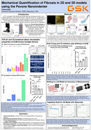
My project for the year was to quantify mechanical properties of fibrosis in 2D ECM models derived
from IPF fibroblasts, using a novel technique and a new instrument called the Pavone Nanoindenter
(Optics11 Life). This technology is based on atomic force microscopy and uses nanoscale indentation
to measure mechanical properties like stiffness and viscoelasticity². I was one of the first people at GSK
to learn how to use this new device and played a key role in setting up and refining the protocols needed
to build capability in this technology. Additionally, my project involved the development of 3D models
of fibrosis utilizing IPF fibroblast spheroids. These are cell aggregates that mimic the architecture and
mechanical environment of human tissue, providing a more physiologically relevant model for testing
anti-fibrotic therapies than generic 2D ECM models.
This report provides an overview of the scientific background of IPF and explains why mechanical
properties were selected as the focus of our study, followed by a description of the experimental design
and execution using the 2D ECM model. I’ll present key findings and discuss their implications, then
outline the early work on setting up 3D spheroid models and their potential value in translational
research. Finally, I’ll reflect the next steps for the project beyond my placement and on the impact I was
able to make during my time at GSK.


Please sign in
If you are a registered user on Laidlaw Scholars Network, please sign in