Week 1 of Research at Leeds: Insights and Reflection
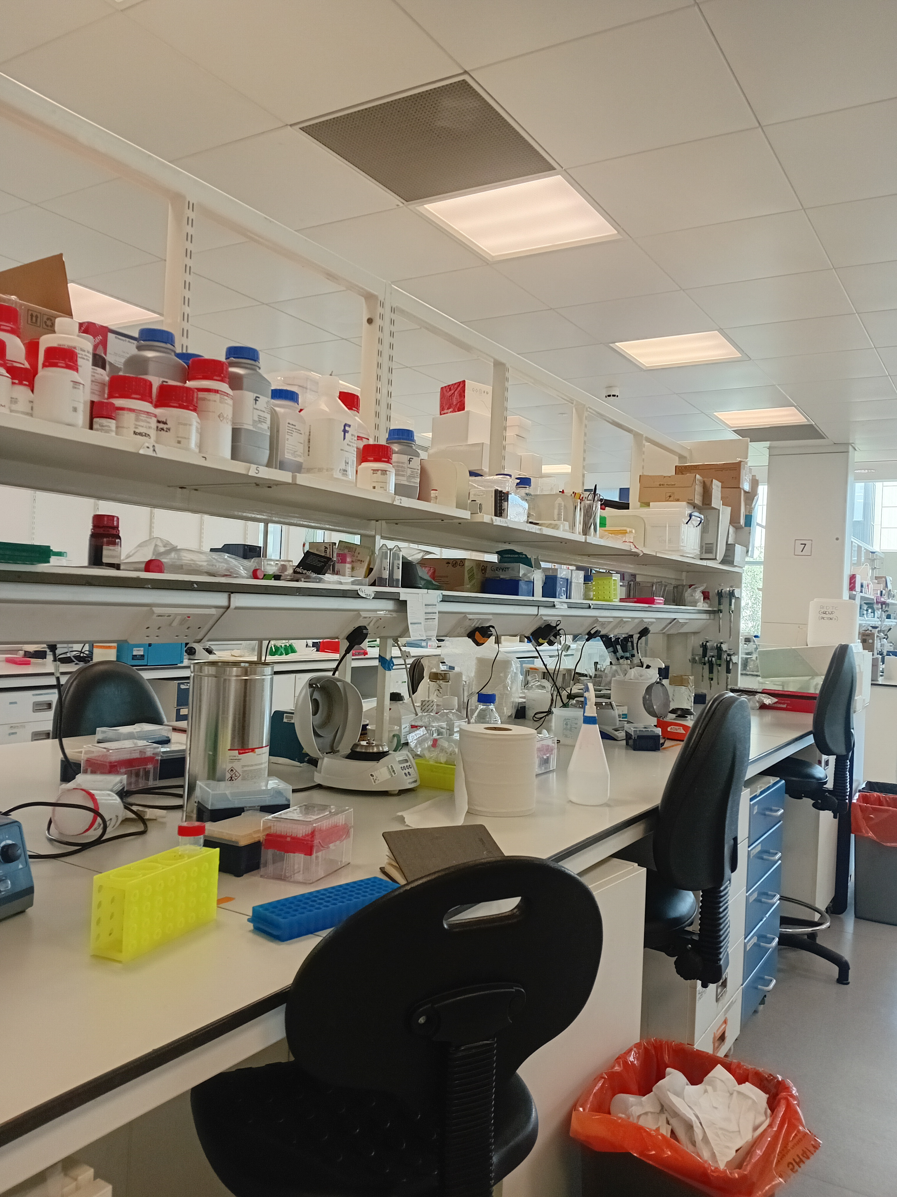
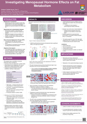
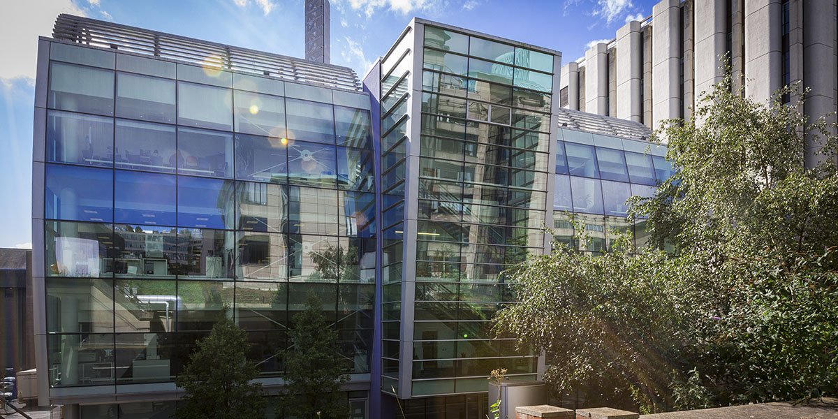
The first week of arriving at Leeds was the most exciting - I did not know what to expect from starting my research placement but felt determined nonetheless to do my best! I got well-acquainted with my supervisor the first day as she showed me around the laboratory and office areas, and it felt surreal to have my presence at the same institute as such impressive scientists and other scholars working on amazing metabolic research at Leeds Institute of Cardiovascular and Metabolic Medicine (LICAMM).
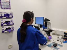
As my entire research project is based on working with mature differentiated adipocytes, I had little room to work on it in the first week when my cells were still in the preadipocyte stage and growing steadily. Nevertheless, it was exciting to check the preadipocyte cells my supervisor had been preparing for me under the microscope. I learned that they had been collected through samples from a liposuction surgery so that we could work on actual female, human cells, undoubtedly making our research premises more realistic (for context, my research is looking into how female adipocytes are affected by change in hormone levels postmenopause). Thus, I spent most of my time acting somewhat like a laboratory assistant for my supervisor, which was incredibly helpful to build many new skills and also get comfortable with the new setting.
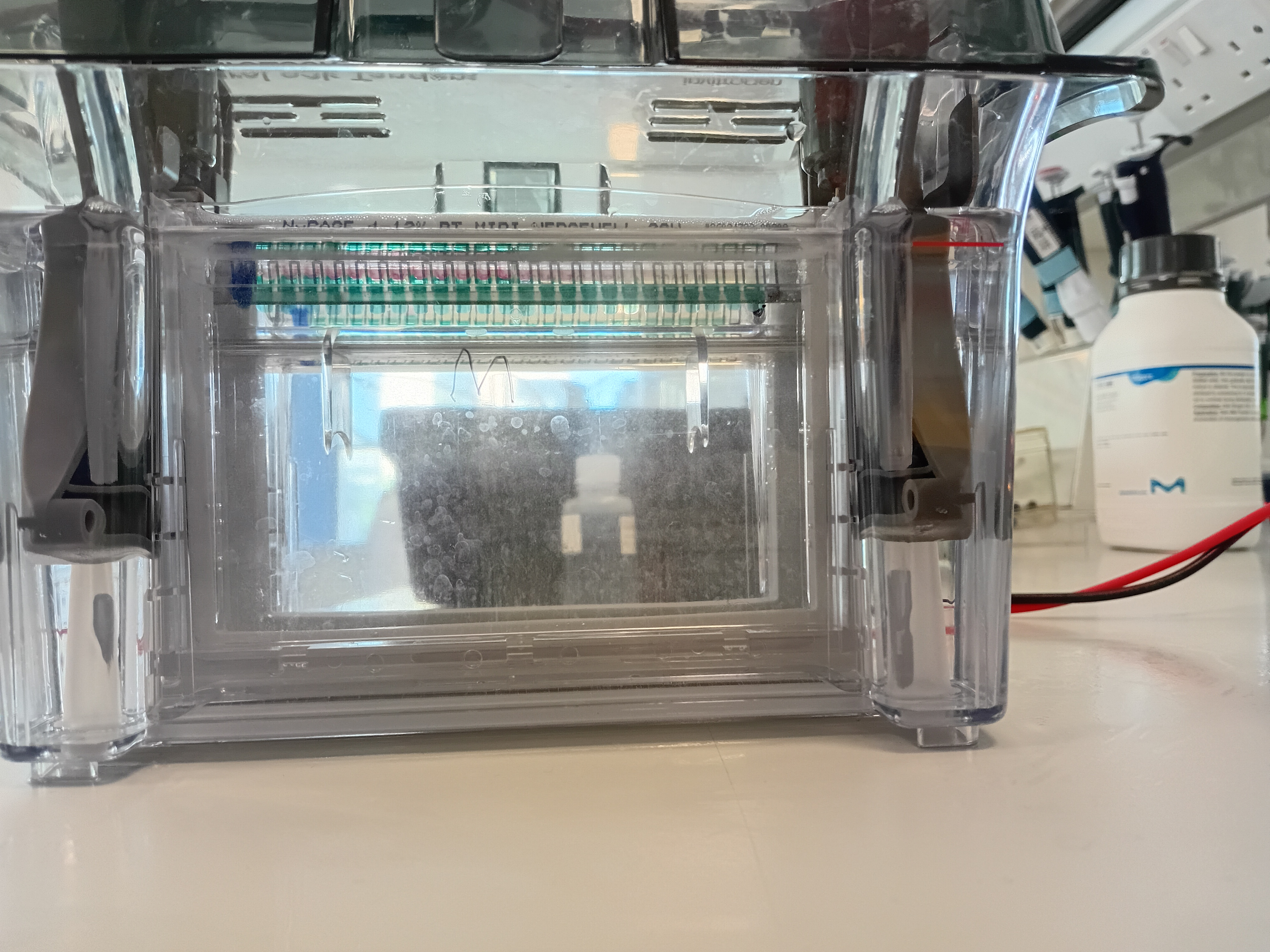
One of the skills I learned and got to practice was carrying out gel electrophoresis, notably loading the wells of the gel, which is definitely harder than it looks since it demands a lot of steadiness and patience. An important step that I was unaware of before is that we must flush the wells with buffer before we start loading the samples as some residue of the gel can impact the results of the electrophoresis! The purpose of gel electrophoresis is to separate a mixture of proteins by size, as smaller proteins find it easier to travel further along the gel.
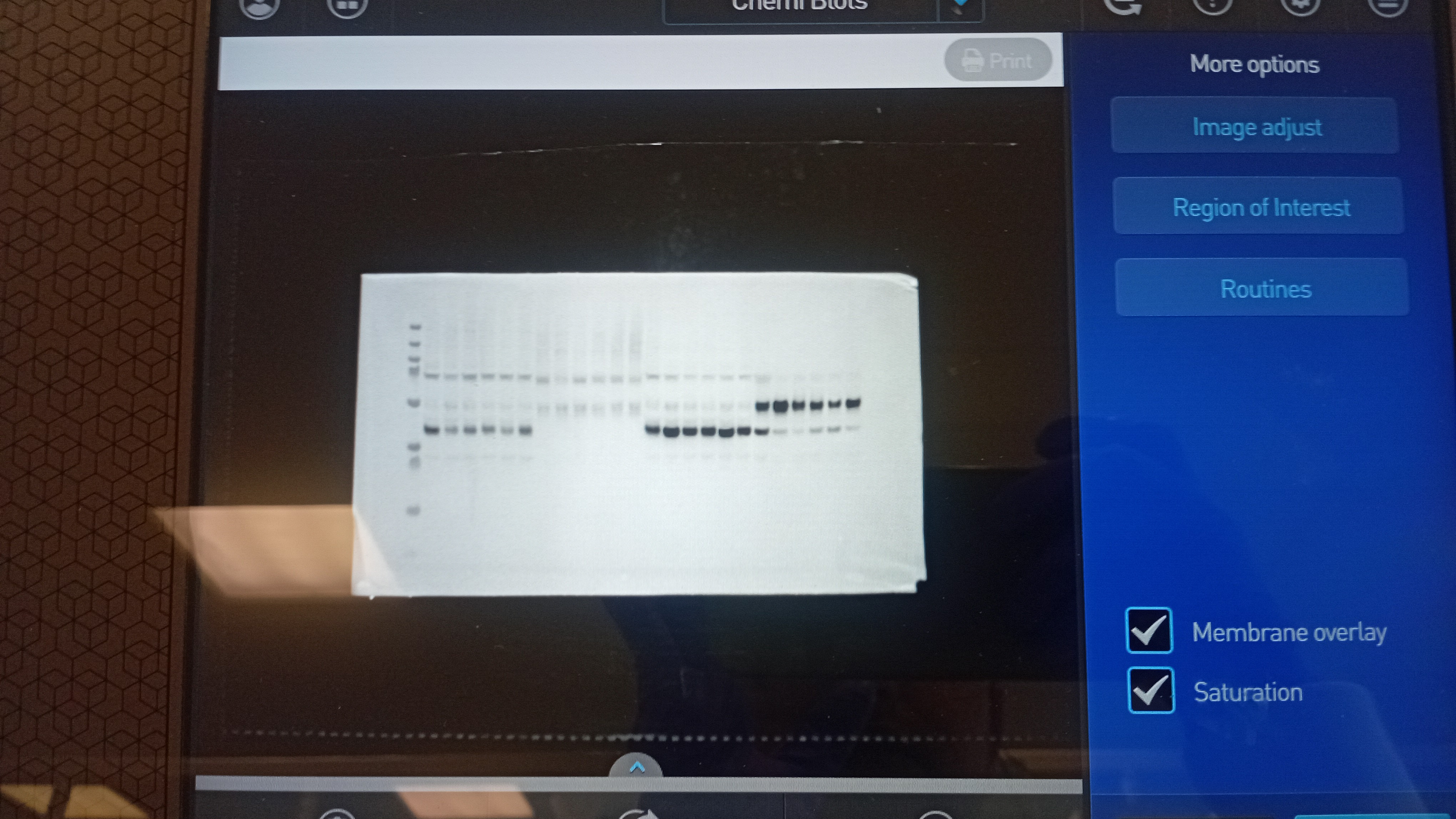
The most important laboratory technique I mastered through repetition and guidance is Western blotting. A western blot visualizes proteins after carrying out gel electrophoresis, and can be used to detect and quantify specific proteins from a complex mixture extracted from cells. Here's how it works: the gel from the electrophoresis is first transferred to a membrane (along with the size ladder), and then the membrane is treated with primary antibodies (that bind to target proteins). The membrane undergoes 5 minute washes with TBS-T 3-4 times on a rocker to get rid of unbound antibodies. Next, a milk wash, also known as blocking, allows milk proteins to add to empty spots on the membrane while secondary antibodies bind to the previous primary antibodies. These secondary antibodies are the ones that can be detected through imaging the blot.
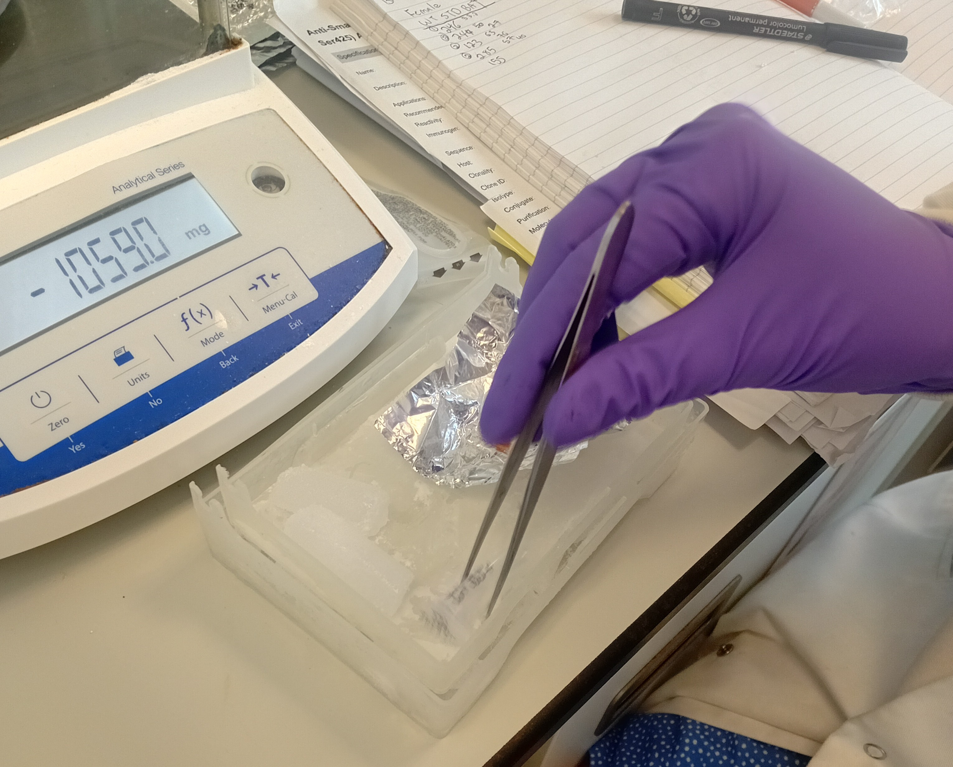
The last skill I acquired is tissue cutting. I helped my supervisor cut some brown adipose tissue acquired from mice and learned a few things about the process. Firstly, the mouse tissue needs to be frozen at all times otherwise molecular changes may be induced in it since chemical reactions can still take place inside the tissue. Thus, dry ice is used to store the tissues and need to be used during the entire cutting process, as well. I used a scalpel and pair of forceps to carefully cut and weigh each piece of tissue, which felt a bit like doing surgery! The cut tissue was later used for protein extraction.
Hi! My name is Naziba and I am from Bangladesh, but currently in Hong Kong for my undergraduate studies at The University of Hong Kong where I study Molecular Biology and Biotechnology.
Besides my academics, I am a very passionate teacher who enjoys making high school science feel more interesting and less taxing. I also love watching movies with my cats and taking nature walks with my friends.
I am excited to start my Laidlaw research journey this year under the Faculty of Health and Medicine at University of Leeds . I am really looking forward to making friends in the Laidlaw community so feel free to reach out in my DMs if you wish to connect!