From Cancer to COVID: Returning to the Bench with the Same Chip but a New Target
Starting my research
In 2023, as I approached the end of my sophomore year of high school, I cold emailed Dr. Shannon Stott, a Principal Investigator at Massachusetts General Hospital. I was part of an Authentic Science Research program at my high school which, across three years, guided me through conducting a literature review, connecting with a research mentor, designing and executing a novel project, and submitting my findings in various formats to high school science research competitions.
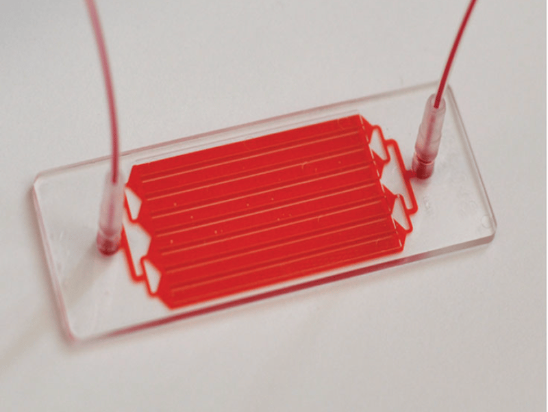
During the summer of 2024, I conducted my research at the Stott Lab, using their microfluidic herringbone chip (HB-Chip) to isolate extracellular vesicles (EVs), membrane bound bundles of RNA, DNA, and protein, from plasma. I then analyzed their contents to determine if these EVs could predict how head and neck squamous cell carcinoma (HNSCC) patients would respond to immunotherapy.
The work was fascinating and rewarding, but also frustrating. Since I was 17, I required constant supervision. Some steps were barred entirely from me due to my age. At times, I felt like a burden to the lab’s day-to-day operations.
This summer, no longer restricted by age-based boundaries and equipped with intricate knowledge of the lab’s equipment and technology, I felt I was in a great position to have a more positive impact and rewarding experience at the Stott Lab.
In addition to working with the Stott Lab, I was co-mentored by Dr. Brian Nahed at Massachusetts General Hospital. As a neurosurgeon who has worked with Dr. Stott in the past, Dr. Nahed offers a unique perspective for how to best utilize liquid biopsy in a clinical setting.
Research background and objectives
Dr. Stott placed me on an ongoing project using the HB-Chip to analyze blood samples from patients with post-acute sequelae of COVID-19 (PASC). Currently, there is no laboratory test to diagnose PASC, commonly referred to as long COVID. The condition, impacting an estimated 36% of confirmed COVID-19 patients, is instead defined as a set of symptoms present for three or more months after SARS-CoV-2 infection. Furthermore, the mechanisms behind PASC are poorly understood. To uncover these mechanisms and work toward a diagnostic test, we processed and compared PASC plasma samples to patients with acute COVID-19, patients who recovered completely after experiencing COVID-19, and healthy controls. In order to ensure the healthy controls had never experienced asymptomatic COVID-19, these blood samples were collected from a bank established before the prevalence of the disease.
Methods
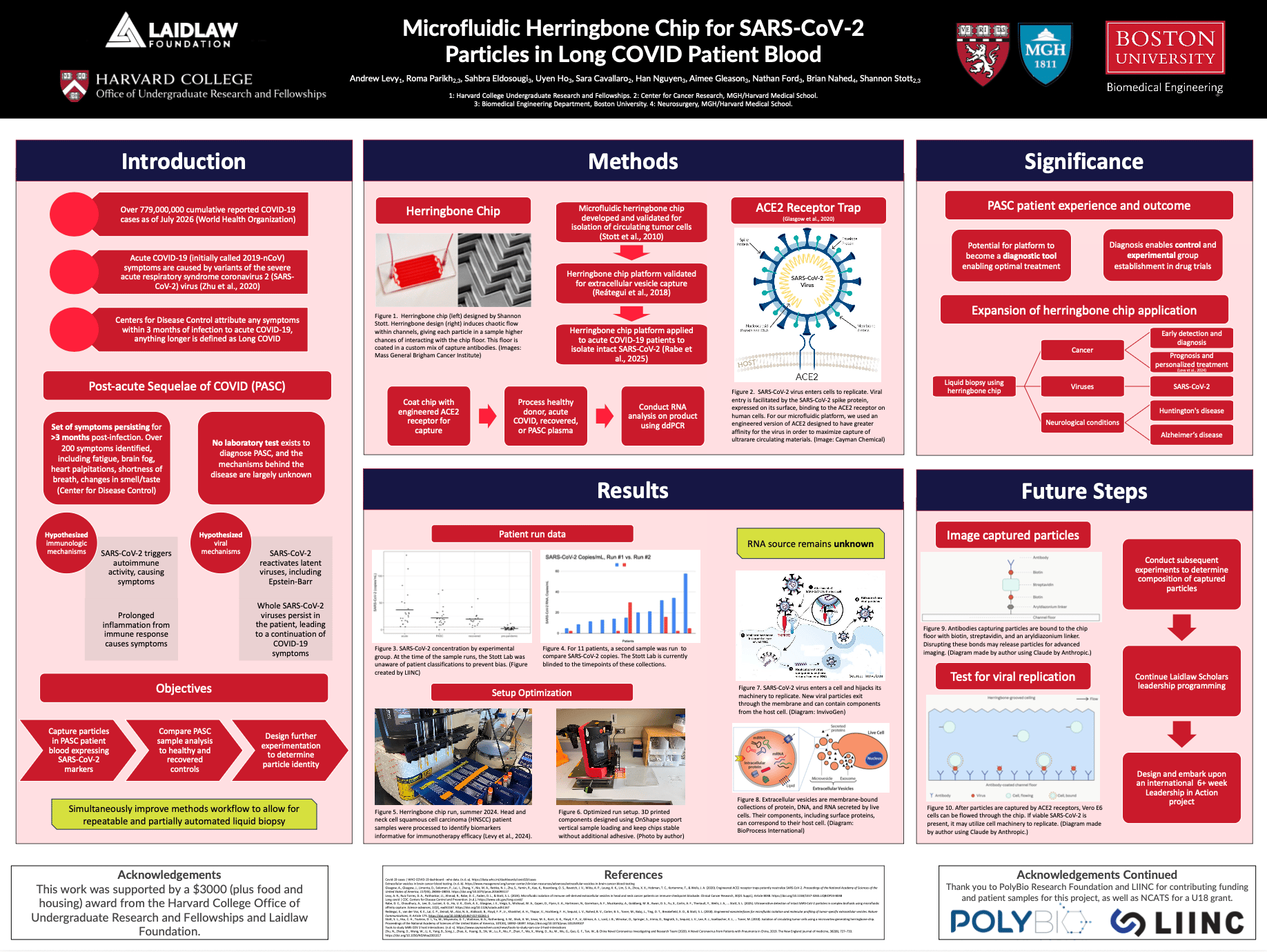
Patient samples were processed with the HB-Chip. This chip has previously been validated for the isolation of cells and extracellular vesicles from cancer patients. Additionally, sample processing can lead to high yield of ultra-rare particles because the chip induces a chaotic flow with its herringbone ridges. This is important because we hypothesized that any viral particles in PASC patients circulated in low quantities.
In order to isolate SARS-CoV-2 materials from patient blood, we leveraged the mechanism by which SARS-CoV-2 enters cells. The HB-Chip has seven channels. Each channel floor can be coated with antibodies through a series of chemical reactions within the chip. First, the chip channels must be functionalized with an aryl-diazonium reaction. Next, streptavidin nanoparticles are pushed through the chip to bind to the floor. Streptavidin binds with strong affinity to biotin. Additionally, most antibodies can be biotinylated, meaning they can be attached to biotin. After biotinylating antibodies, we push them through chips already coated in the streptavidin nanoparticles. Thus, the antibodies bind to the floor of the chip, enabling particle capture.
Importantly, the antibodies the HB-Chip is coated with can be customized depending on the intention of the assay. For my previous work exploring the immune environment in patients with HNSCC, I utilized antibodies with demonstrated affinity to B cells (CD19 and CD20). For this project, we coated the chip in an engineered ACE2 receptor that binds to viral spike proteins. This receptor was optimized to ensure heightened affinity to SARS-CoV-2.
Once the HB-Chip floors were coated with ACE2, we conducted the processing of patient blood samples. Our protocol was as follows: Plasma samples were thawed from storage at -80℃ and loaded into plastic syringes. Syringes were fitted with needles. Needles were fed into plastic tubing, connecting the syringe to the chip inlet. Syringes were then loaded into automatic syringe pumps, which are able to maintain an exact and automatic flow rate of sample through the chip (1 mL/hour). These samples were then pushed through the chip.
As plasma flowed through the HB-Chips, we hypothesized that viral particles in the bloodstream would be captured by the ACE2 antibodies coating the floor of the chip. Thus, membrane bound bundles of RNA would be present on the chip. After a wash step, where phosphate buffered saline (PBS) was run through the platform, we hypothesized that these viral particles remained on the chip.
In order to confirm the presence of viral RNA within membrane-bound particles, we needed to free the RNA from the chip. Thus, we conducted a lysis step to dissolve any membranes present. We used a pre-established lysis solution and pushed it back and forth through the chip at set flow rates. However, here we ran into an inconvenience. This step demanded the syringes be loaded vertically, but the syringe pump is designed for horizontal use. Thus, when the pump is placed on its side, it is not balanced. Additionally, spacing of chips can be inconsistent since they are held in place by nothing but labeling tape. We required both a method to secure the pump and a method to secure the chips. We approached these problems with the same potential solution. Using OnShape, a computer-aided design (CAD) program, we designed a stand to hold the syringe pump and a tray to secure the chips. Once printed, these devices drastically improved our workflow. Our chance of error (via a pump falling or chip sliding) decreased, and the system became easier to set up. The results of these improvements can be seen in the results section of my poster (attached).
After performing the lysis step, leaving us with RNA suspended in a buffer solution, we now had to identify the genetic sequences discovered. To do so, we analyzed these RNA samples with droplet digital PCR (ddPCR). ddPCR serves a different purpose from RNA sequencing. RNA sequencing is a more expensive procedure that reveals the full genetic sequence of the segment. ddPCR packages individual RNA segments into oil droplets and uses fluorescence detection to determine the frequency at which the segments match a predetermined segment. In other words, RNA sequencing maps the RNA while ddPCR quantifies the presence of a preestablished segment.
Results
Our results consisted of two steps: Initial runs and secondary runs at a blinded timepoint. Figures for both are shown on my research poster (attached). These figures were created by the Long-term Impact of Infection with Novel Coronavirus (LIINC) study, who generously provided samples for this project. Unfortunately, since this research is ongoing and the Stott Lab is blinded to the identifications of the patients, we were unable to conduct further statistical analysis.
However, we did find a clear difference between healthy pre-pandemic donors and all other patient groups. These other patient groups included recovered COVID-19 patients and PASC patients. This demonstrates that SARS-CoV-2 particles likely continue to circulate in patient bloodstreams for months after infection. One of our project goals was to work to define the mechanism causing PASC. While our evidence is not definitive, it provides a basis to explore the existing theory that SARS-CoV-2 particles persist in patients and cause symptoms. However, our results do not invalidate other existing theories, including that PASC is caused by autoimmune activity, prolonged inflammation, or reactivation of other latent viruses.
We expect in our continuing analyses to observe greater differences in RNA concentrations among our patient groups. This would validate the pursuit of a blood-based diagnostic test for long COVID. Establishing a definitive diagnostic benchmark for long COVID would enable exploration and testing of treatment methods, improving patient quality of life.
Future research steps
While we were able to capture membrane-bound viral RNA, we are unsure of the identity of these viral particles. We have two hypotheses. The first is that they are hybridized viruses/EVs. If SARS-CoV-2 RNA remains latent in cells after infection, their RNA and proteins may be present in EVs released by cells. The second is that these particles are SARS-CoV-2 whole viruses. Differentiating between these two theories is important to explore the future applications of our platform and the impact of our results. However, given that EVs and viruses share similar qualities (membrane-bound bundles of RNA expressing spike proteins that can be roughly 100 nm in diameter), this differentiation is difficult. Thus, I was tasked with designing future experiments to differentiate between these outcomes.
One experimental design plans to disrupt the bonds between captured components of the blood and the chip, freeing the particle. Subsequently, cryogenic electron microscopy (cryo-EM) can be used to image individual particles. Our hypothesis is that hybridized EVs would visually contain fewer spike proteins compared to intact SARS-CoV-2. By comparing images of our captured particles to existing images of SARS-CoV-2, we may be able to determine whether we have captured the whole virus.
However, this experimental design has a critical limitation. The HB-Chip bonds were designed to act with maximal strength and affinity to capture rare circulating particles. Thus, these bonds are difficult to disrupt. Ongoing Stott Lab research is testing different buffers’ ability to disrupt these bonds. The most promising approach is to vary the buffer pH, which could create instability in chemical bonding.
Another experimental design would seek to test the ability of captured particles to undergo processes normal for intact SARS-CoV-2. While EVs are created in large quantities by host cells, they cannot replicate on their own. However, SARS-CoV-2 is able to infiltrate cells and utilize their machinery to replicate and proliferate. I hypothesize that, without removing captured particles from the HB-Chip, we can evaluate their ability to replicate. The Vero E6 cell line has been validated as a host for SARS-CoV-2 replication in vitro. Thus, if Vero E6 cells are pushed through the chip after viral particle capture, and whole viruses are present, the viruses may be able to replicate. This could result in a thousandfold increase in whole virus concentration, causing a higher ddPCR readout. Additionally, since replicated viruses in cells eventually cause cell membrane lysis, we could quantify particle count without conducting a lysis step. If we observe such evidence of replication, it would support the hypothesis that we are capturing intact SARS-CoV-2 with the HB-Chip.
Significance
Our primary study goals were to improve PASC understanding and to work toward a diagnostic platform. However, the significance of this research has a broader impact.
As previously mentioned, the HB-Chip can be applied to a variety of diseases and conditions. Before the COVID-19 pandemic, the Stott Lab had focused its resources on cancer diagnosis and prognosis through the isolation of EVs. The application of EV-based diagnostics however is far more vast. Neurological conditions including Parkinson’s disease and Alzheimer’s disease may present with altered blood composition. By determining if unique EVs exist in the blood of these patients, we may gain the ability to utilize the HB-Chip as an early screening device for such diseases.
Importantly, our work with SARS-CoV-2 demonstrates that the capture of viral particles is possible with the HB-Chip. The versatility of our platform via the customization of capture antibodies could allow us to adapt the HB-Chip for the diagnosis of other viruses.
Overall, the HB-Chip proves to be an effective platform for capturing circulating particles. This ability may be valuable in a variety of clinical applications. Our research validates continuing efforts to increase the versatility of the HB-Chip and its use for other clinical challenges.
My future role
My presence at the lab was particularly rewarding this summer. The Stott Lab underwent a change in lab space from Massachusetts General Hospital to Boston University. Collectively, our lab had to move, unpack, and label equipment in new spaces. This was a valuable experience. I was taught organizational skills and the value of spending extra time on little improvements that make experimentation easier.
However, this change also meant my wet lab workload was not as high as I anticipated. Thus, I aim to continue my research efforts with the Stott Lab. We will carry out proposed future steps while simultaneously expanding the application of the HB-Chip for other patient populations.
For my Laidlaw Leadership in Action project, I am considering multiple directions. One goal I have is to expand HB-Chip application for research, diagnosis, and prognosis. There is a lab in Oxford, England, working to apply the HB-Chip to emergency room triage. This research goal is of particular interest to me given my eventual goal of working as an emergency physician. Additionally, a lab in Rome, Italy, is working to apply the HB-Chip to pancreatic ductal adenocarcinoma (PDAC). Working with these labs to improve their HB-Chip workflow would create a positive impact on researchers and patient populations abroad. Another goal I have is to increase the preparedness of healthcare systems to combat pandemics. There are a number of international nonprofit organizations that work to improve healthcare systems through education and resource dispersion. Working with one would be an honor.
In any case, I look forward to translating my research efforts into concrete steps to improve patient livelihoods. I am grateful to the Laidlaw Foundation for granting me this opportunity.
Acknowledgements
I’d like to thank Dr. Shannon Stott, Dr. Brian Nahed, Dr. Roma Parikh, Sahbra Eldosougi, Uyen Ho, Dr. Sara Cavallaro, Han Nguyen, Aimee Gleason, and Nathan Ford for their tremendous support in the lab. I would also like to thank PolyBio Research Foundation and the LIINC study for providing funding and samples for this project.
My involvement and lab impact would not have been possible without Matt DelSesto, who has led the inaugural Harvard Laidlaw Scholars cohort through Harvard Undergraduate Research and Fellowships. His support and guidance has been invaluable. I also want to thank the 14 other members of Harvard’s inaugural Laidlaw Scholars cohort, who have created a culture of support, collaboration, and community that made my experience fulfilling.
I’d also like to thank my parents for their constant support and guidance. They make me feel like I can do anything. I also want to thank my brother for making me a better person, and my sister for inspiring me to pursue both research and medicine. She is my biggest inspiration and role model.
Research Poster