At the Scalpel's Edge - Week 1
Today marks the end of the first week of my Laidlaw research project. There was a lot that was accomplished and learned, one of the things being the importance of the fine motor control of the hands when dissecting specimens.
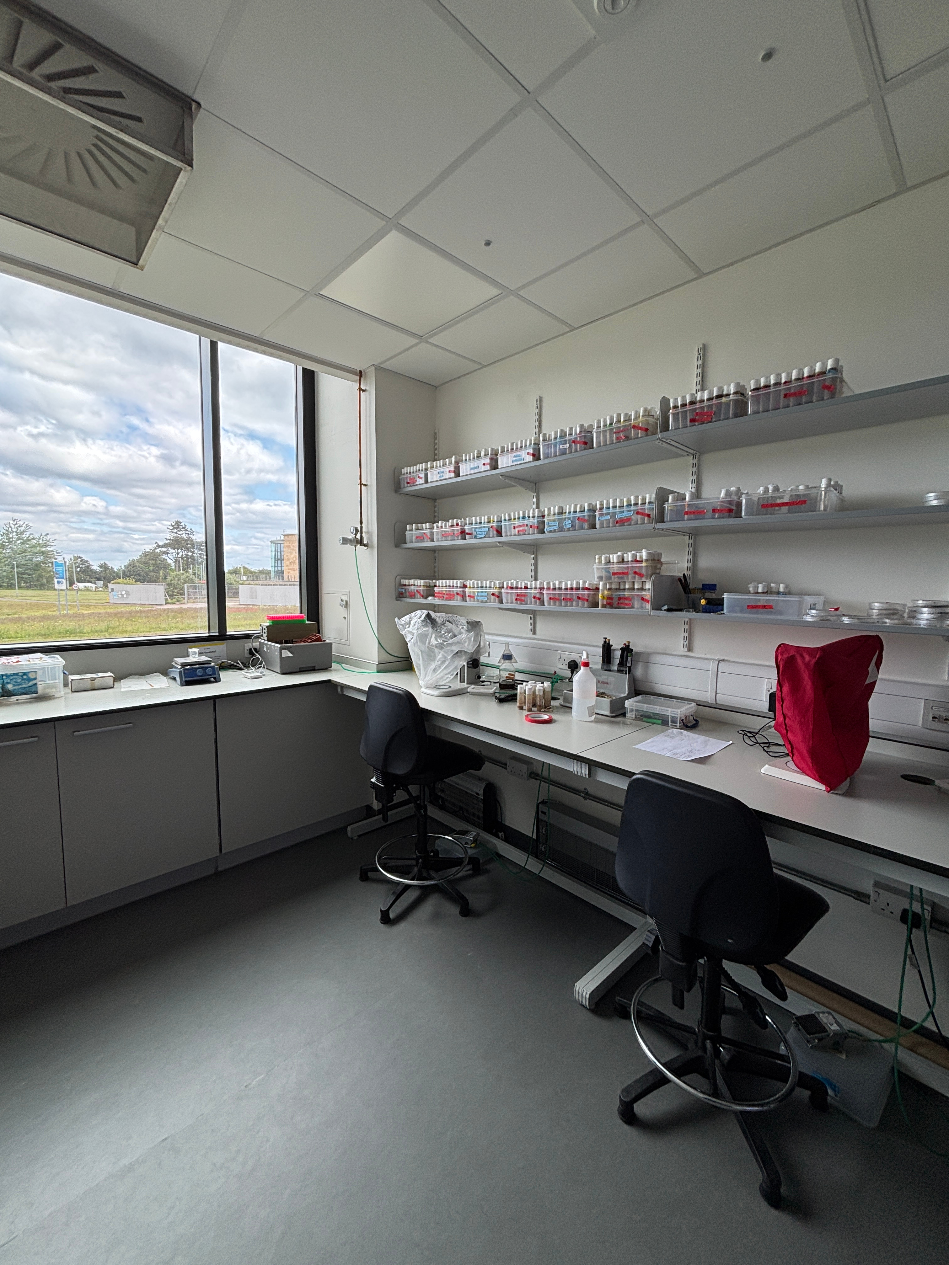
My first three days, namely Monday, Tuesday, and Wednesday, were spent practicing dissection on Drosophila melanogaster flies. The days followed the same old, dull routine: coming into the lab in the morning, going into the fly room, putting on a lab coat, setting up slides, choosing pupae from fly tubes, fixing them on the tape with tweezers, transporting the slides to a separate lab room, and proceeding to dissect them using a scalpel. Thus, memorising song lyrics, such as those from Escape (The Piña Colada Song, that I could hear oscillating from the radio in one of the labs was a way to boost morale when I spent an hour dissecting 30 flies only for none of them to be suitable for staining.
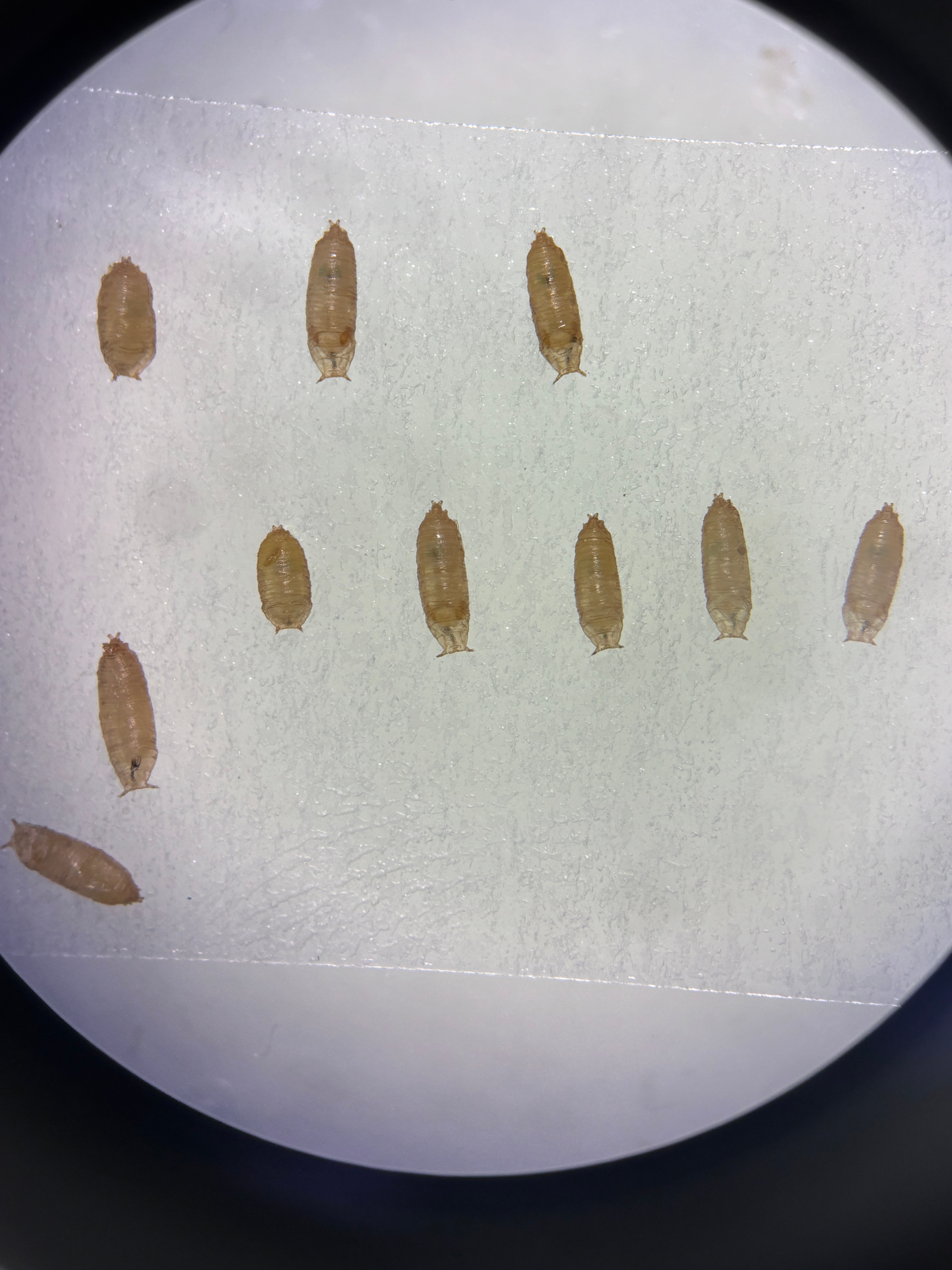 Figure 1: D. melanogaster pupae on a glass slide.
Absorbed by my lab work, my only excuse to go outside for a small chunk of time, which I tried to stretch out for as long as possible, was to sift through dirt obtained from potted plants in the write-up office area. Despite my best efforts to enjoy the fresh breeze that had come to visit me from the North Sea, the weather did not make it easy. Nonetheless, I was able to relive my childhood memories of playing in the sandpit, only now the sandpit could fit on my lap, it was dirt and not sand, and my mission was to find small pupae instead of building castles.
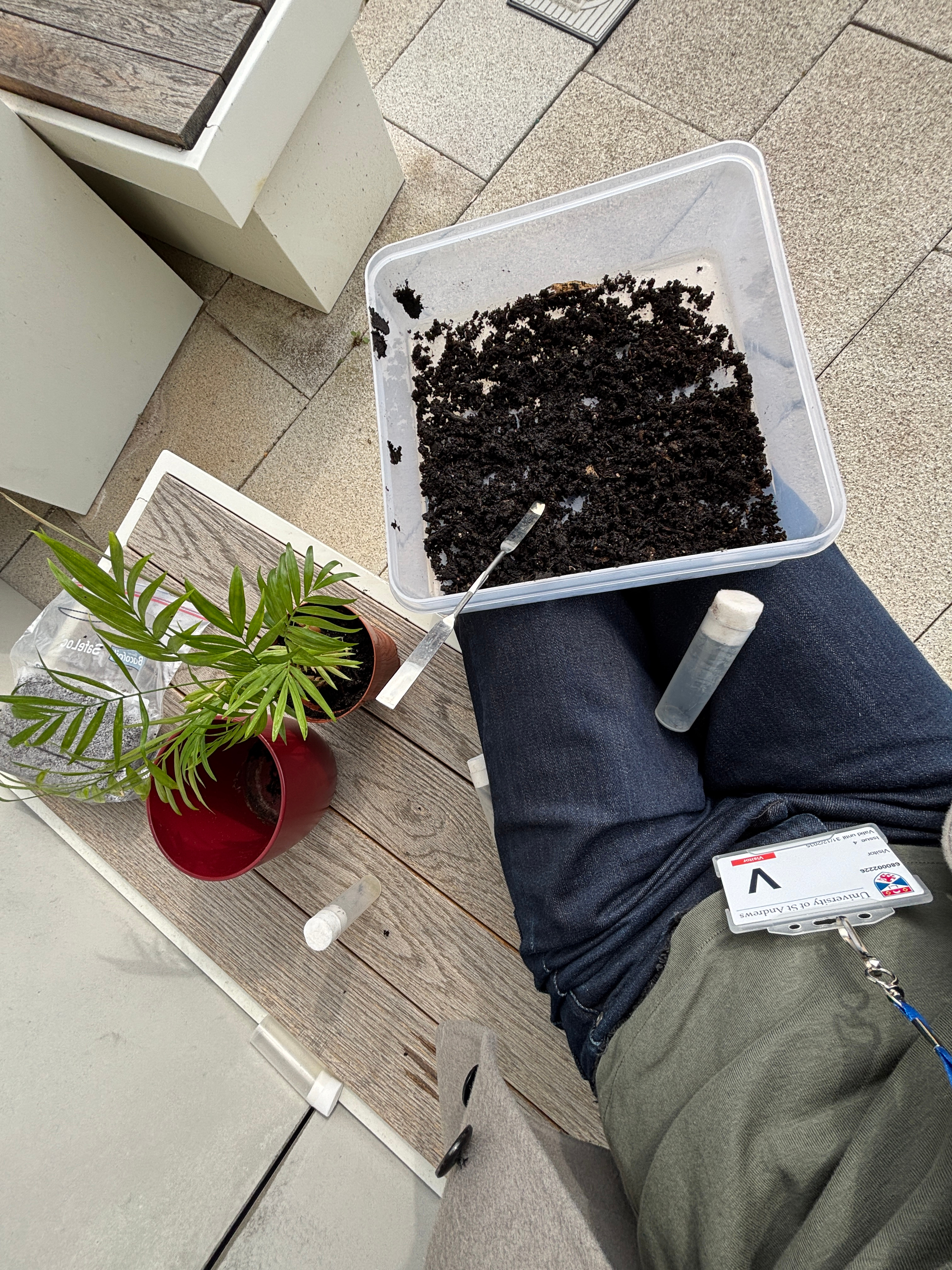 Figure 2: In search of suspected Bradysia sp. fungus gnat pupae, larvae, and/or adults in the soil collected from plant pots.
When Thursday came around, my supervisor and I got on a train headed to Edinburgh as a part of our mission to collect three batches of Anopheles sp. mosquitoes from the University of Edinburgh. This seemed simple: transport mosquitoes from Lab A, Lab A being a malaria lab, to Lab B. However, it is not until I found out how we were going to transport them that the challenge became clear. Mosquito larvae and pupae are aquatic, therefore, they must be transported in water. My supervisor's solution was to put them into ice cream containers, wrap them from end to end in tape, and put the said containers into two bags from Tesco. As he put the last container into the bag, and looked at me, I realised that my role would be to carry them all the way back without any leakages. I, being very clumsy, was quite hesitant but, I took my job seriously. In spite of a few sploshing sounds during moments of turbulence, the containers arrived safely to our lab back in St Andrews.
The real work began on Friday and spilled into Saturday and Sunday. I spent all my time adapting to cutting the soft bodies of the larvae and the pupae that did not have a pupal case (unlike the D. melanogaster pupae which I had practiced on). Once I had cut the larvae and pupae, I stained them and then proceeded with the visualization of the cut specimens using a confocal microscope. Unfortunately, none of the images produced can be used in the final essay; however, the photographs of the specimens helped me understand how to improve my dissection and learn the morphology of the specimens (and, they were so pretty to look at!).
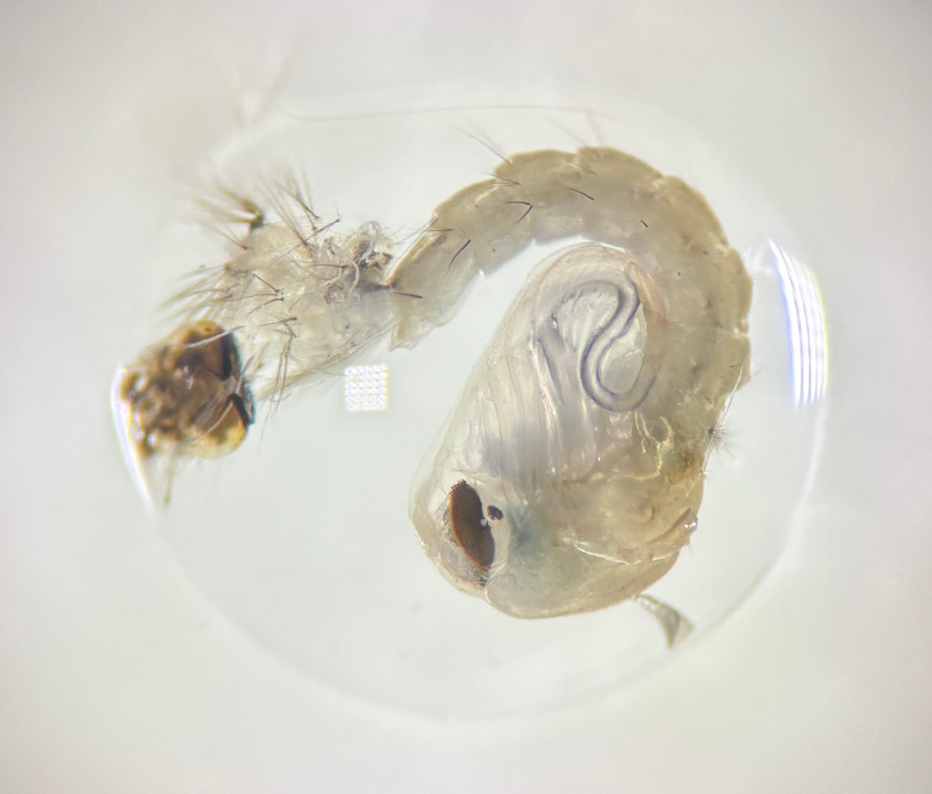
Figure 2: Anopheles sp. mosquito pupa that has shed its larval molt. The photo was taken using a light microscope on an iPhone camera.
On such busy days, it was difficult to have a moment to reflect on my growth, as after the lab I would immediately rush to make dinner and catch up with my fellow Laidlaw Scholars over a cup of tea. However, now as I am writing this on a more quiet evening before heading off to bed, I can see that I already feel more confident as a researcher. I practiced keeping a lab notebook, gained enough experience to dissect the pupae and larvae specimens, understood the conditions needed for the Hoechst staining, and became more familiar with identifying morphological structures. Furthermore, I attended an enlightening group meeting where I learned what research is currently being carried out by a Master's student in the same lab on the membrane microdomains present in D. melanogaster. (Compared to the important progress of the PhD student, my findings paled in comparison: I found that it is possible to culture a new population of fungus gnats (which might potentially also be used in my research) using honey.)
My goals for next week are to find several larvae directly before they turn into a pupae, continue working on improving my dissection so as to preserve as much of the tissue as possible, and start working on understanding the timing of development to be able to produce time-accurate results.
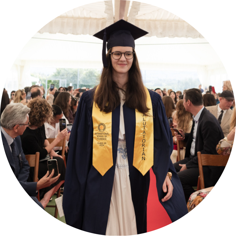
Hello hello! I am an incoming fourth-year student at the University of St Andrews pursuing a degree in biology. Originally from Kyiv, Ukraine, I moved to Italy in 2013 and have been living there ever since.
Over the course of my academic journey I have had the chance to explore numerous areas in biology, ranging from evolution to bacteriophage discovery to cell systems, out of which epigenetics and gene regulation during development have piqued my interest.
My primary research interest is developmental biology, which arose from my fascination with how multicellular organisms originate from a single cell. Therefore, my research focus for the first summer of the Laidlaw Scholars programme was the investigation of abdominal metamorphosis in Dipteran insects. To do so, I used fixation and Hoechst staining in addition to microscopy to study cell movement during several developmental stages in 2 species of flies.
As for my leadership in action (LiA) project, I decided to assist biodiversity monitoring and conservation efforts in the heart of the Amazon rainforest jungle in Peru.
If I am not in lectures or studying in the library, I can be found working on a short story, watching films (and logging them on Letterboxd), reading old science fiction, or on a hike capturing the nature around me through photography. Having grown up in a multicultural setting, I enjoy learning more about the different cultures that exist on our planet, whether it is through cuisine, music, travelling, or linguistics. The latter partially explains my grasp of nearly five languages, though I have to warn you that my fluency differs dramatically. Otherwise, I also love spending time with animals, playing board games, or just talking.
More than anything, I am always happy to meet like-minded individuals who are curious about the world. Thus, please feel free to reach out or connect with me on LinkedIn!